

What To Do For A Nail Avulsion
Department of Dermatology and STD, Academy College of Medical Sciences and Associated Guru Teg Bahadur Infirmary, Academy of Delhi, Delhi, India
Correspondence Accost:
Deepika Pandhi
Department of Dermatology and STD, University College of Medical Sciences and Associated Guru Teg Bahadur Hospital, University of Delhi, Delhi 110 095
Bharat
Introduction
Nail avulsion, the separation of the nail plate from the surrounding structures, is the most frequently performed surgical or nonsurgical, chemical procedure on the nail unit. It may either be useful to explore the blast unit for diagnostic purposes or as a therapeutic tool in particular nail pathologies. Boom avulsion may be accomplished using either a distal or a proximal anatomical approach. The former is the most often used technique, in which the nail plate is released from its attachment from the nail bed at the hyponychium, [1] while in the latter, the blast plate is separated from the proximal nail fold (PNF) followed by a complete separation moving distally. [1] Chemic nail avulsion past using urea ointments is a valuable technique that avoids the complications encountered with its surgical counterpart. The current dissertation is an effort to bring nearly the salient briefs of the discipline succinctly.
Applied Anatomy
It is imperative to understand the anatomy of the nail and the intricacies of nail growth prior to undertaking nail avulsion. Nails are ectodermal appendages covering the dorsal aspects of the digits. [2]
These structures provide protection and integrity to the fingertip, in add-on to facilitating skilled hand functions. Therefore, an aberrant alteration in the anatomy of the nail unit may interfere with the aforementioned functions. The distal border of the nail is free, while the proximal edge is clutched into a fold and is covered with a skin flap called the eponychium or cuticle [Figure - 1]. The nail is attached at its lateral, distal and proximal borders. The nail bed, also called the sterile matrix, anchors the dermis to the periosteum of the distal phalanx. The matrix has been divided into distal sterile matrix (nail bed), which is covered with grown blast, intermediate matrix that corresponds to the epithelial lining of the ventral surface of the PNF, and the distal germinal matrix, from which a new nail arises. The germinal matrix is covered with the eponychium. [2]
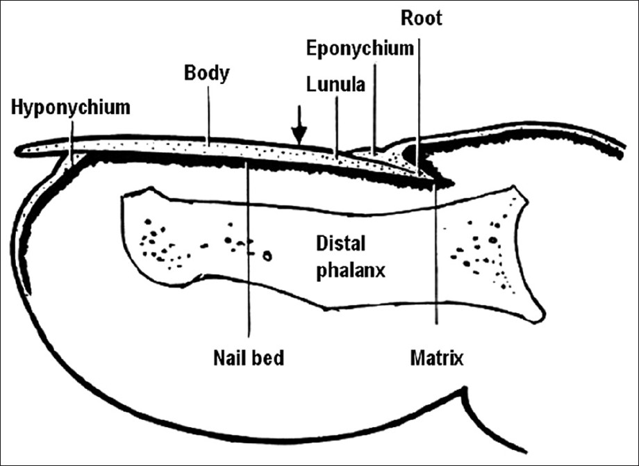 |
| Figure one: Anatomy of nail |
The lunula is the pale crescent-shaped structure easily recognized under the proximal portion of the nail. Importantly, the blast is not firmly attached at the lunula. Considering the blast is formed in the germinal matrix, loss or deformity of this role results in permanent loss or permanent deformity of the boom. As the boom grows distally, the superficial cells become cornified. Distal to the lunula, the nail is firmly fastened to the nail bed or sterile matrix. [2] Consummate regrowth of an avulsed finger nail usually requires 4-v months (1 mm/calendar week), whereas the toe nail may require up to ten-12 months. Information technology is essential to preserve the peel folds surrounding the smash margins. Broad scars or misalignment in the peel fold can issue in splitting or permanent deformity of the boom when it regrows. Adhesions betwixt the eponychium, nail bed and matrix are prevented by maintaining this space with either the replaced smash or gauze packing. The skin of the nail bed is supported by a highly vascularized subcutaneous layer that intimately links the blast bed to the dorsal periosteum of the distal phalanx. [3] The subungual glomus is a rich vascular network of microscopic vessels situated in the subcutaneous tissue deep to the nail bed, and plays a role in peripheral temperature regulation. Distal to the hyponychium and plantar to the medial and lateral nail folds (LNF), the digital pulp surrounds the distal phalanx and conveys vessels and fretfulness to and from the toe tip.
Indications of Smash Avulsion
Nail avulsion is the nigh common surgical procedure performed on the nail unit. The nail plate is excised from its prime attachments, the nail bed ventrally and the PNF dorsally. The indications of nail avulsion are outlined as follows:
Diagnostic
Blast avulsion is oft undertaken every bit a preliminary step for the following indications: [4]
- Exploration of the nail bed and the nail matrix: This may exist required in order to look for the pathologies originating in either the blast bed or the nail matrix, which include inflammatory dermatoses, infections, connective tissue diseases and tumors. On the other paw, a disease process affecting the surrounding tissues may encroach on the nail bed.
- Exploration of the PNF and the LNF: A complete exposure of these structures to divulge the extent of a disease may require a nail avulsion.
- Performing biopsy on the smash bed and the nail matrix: Many a times, nail avulsion is performed to uncover the nail bed and matrix for the purpose of a biopsy. This is frequently the situation in diseases like psoriasis, lichen planus, twenty nail dystrophy, nail unit tumors, nevi, melanonychia and pachyonychia congenita.
Therapeutic
Other than the aforementioned indications, nail avulsion is used every bit a therapeutic adjunct for the post-obit indications:
- Before a chemical or surgical matricectomy: Matricectomy refers to the complete extirpation of the nail matrix, resulting in permanent nail loss. Unremarkably, notwithstanding, matricectomy is simply fractional, restricted to one or both lateral horns of the matrix. Nail ablation is the definitive removal of the entire boom organ. The near important mutual denominator in a successful matricectomy is the full removal or devastation of the matrix tissue. Matricectomy may exist indicated for the management of onychauxis, onychogryphosis, congenital nail dystrophies and chronic painful nail, such as recalcitrant ingrown toenail or split within the medial or lateral one-tertiary of the nail.
- Ingrown toe blast/onychocryptosis: Indications for the handling of an ingrown toenail include significant pain or infection, onychogryphosis (a plain-featured and curved nail) or chronic, recurrent paronychia (inflammation of the boom fold). The most mutual procedure to treat locally infected ingrown toenails is partial avulsion of the lateral edge of the nail followed by chemical matricectomy using eighty-88% phenol (phenolization). [5],[half dozen] In a randomized report [vii] comprising 117 patients, patients underwent fractional blast avulsion in combination with either excision of the matrix or application of phenol, with or without local awarding of gentamicin afterward. The measured endpoints were infection at ane week and recurrence at ane year. Infection rates were found unrelated to the use of antibiotics. Yet, recurrence rates were found to be significantly lower after phenolization of the smash bed (xiii.eight%) compared with excision of the boom matrix (38.9%). Contrary to this, another randomized trial comprising 63 patients institute both partial smash avulsion with phenolisation or with partial matricectomy to exist equally effective. [8] As well, a remarkably low incidence of recurrence (0.half dozen%) and wound infection (two%) has been found. The hateful time to render to normal activities is ii.1 weeks. [9]
- Chronic onychomycosis: Xiii patients with distal subungual onychomycosis in a total of 48 dermatophyte-infected nails were treated with chemomechanical, fractional nail avulsion followed past topical miconazole for 8 weeks. Periungual pare irritation was common during the initial avulsion period. The clinical and mycological cure rate was 42% at 6 months afterward cessation of therapy. The therapeutic response was related to the pretreatment extension of subungual hyperkeratosis. This treatment modality could be a valuable alternative to other remedies for the handling of onychomycosis limited to a few nails. [10] Full nail avulsion has been found to be constructive, especially for patients with single- or oligo-onychomycosis [Figure - 2] and in those with a dubious diagnosis. [11] In dissimilarity, some other randomized trial comprising 40 subjects with single-smash onychomycosis recorded a high drop-out rate. All cases of total dystrophic onychomycosis failed to respond to this therapy. Overall, 15 of 27 (56%) patients were cured with this arroyo. No side-effects or long-term complications of the boom avulsion were encountered. [12]
-
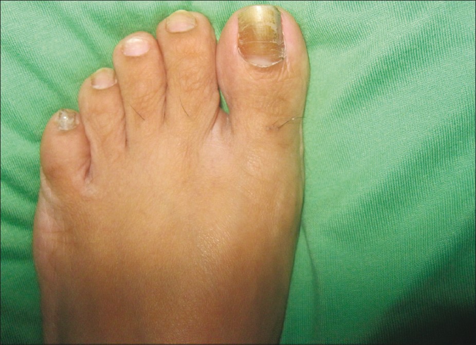
Figure ii: Distal subungual onychomycosis involving a single blast with evidence of onychogryphosis - Traumatic nail injuries: Avulsion may be used to evaluate the stability of the nail bed or to release a subungual hematoma afterward failed puncture aspiration. If sufficient blunt or sharp force is applied to the nail plate and surrounding folds, it can violate the structural integrity of the blast bed and the resultant hemorrhage can fill the potential space that unremarkably exists between the nail plate and the underlying smash bed. The force of the injury as well as the hemorrhagic response tin separate the nail plate from the bed, causing traumatic onycholysis. If the force is sufficient plenty, the proximal margin of the plate will ofttimes divide from the matrix region nether the PNF and elevate through the nail fold. This disrupts the seal of the cuticle and potentially exposes the underlying tissues to bacterial contamination. If there is an associated fracture, the patient may exist at risk for distal phalangeal osteomyelitis. [13] Whenever a patient presents with an acutely injured, throbbing toe with a subungual hematoma, one should consider disruption of the nail plate. If the patient maintains structural integrity of the smash folds and there is disruption of the nail bed, subungual force per unit area secondary to hemorrhage tin cause persistent digital pain that may final for several hours to several days, and merely draining the hematoma will usually provide relief. There are many ways to drain a painful subungual hematoma safely. The method i uses is based on the structural integrity of the nail folds and the amount of the visible nail plate associated with the hematoma. Every bit a dominion, when there are stable nail folds and an injury displaying less than 25% of the visible nail plate associated with the hematoma, one tin drain the hematoma through the nail plate. [14] If the subungual hematoma involves greater than 25% of the visible nail plate and/or the boom plate has been avulsed in such a way every bit to disrupt the proximal, medial or LNF contiguous with the bed, then a significant nail bed laceration is probable. Appropriately, 1 should remove the entire plate in social club to facilitate straight visualization and surgical repair of the nail bed. Astringent stabbing or plantar flexor injuries tin can cause boom bed laceration and phalangeal fractures that propagate along the dorsal surface of the nail plate into the PNF and through the physeal plate of the distal phalanx, separating the nail plate from the ventral surface of the PNF. [fifteen] When this occurs, the basilar epiphysis is usually displaced dorsal relative to the nail bed because the epiphysis remains anchored to the interphalangeal collateral ligaments and the extensor tendon. [16] Ane would treat this injury by removing the nail plate or at least the proximal portion of the nail plate and post-obit-up with cleansing, debridement and inspection.
- Chronic paronychia: It is an extremely recalcitrant dermatosis that is particularly prevalent in housewives. Medical treatment for this condition is unsatisfactory in a significant number of cases. [17] In these patients, no response is evident to irritant avoidance and topical therapy and surgical approach forms a vital part of management. An en bloc excision of the PNF combined with a total, or more normally fractional, restricted to the base of the nail plate, smash plate avulsion has been shown to exist a useful method in chronic, recalcitrant paronychia, specially where the PNF is fibrosed or thickened. [17] Alternatively, an eponychial marsupialization, with or without blast removal, may be performed. This technique involves excision of a semicircular skin section proximal to the nail fold and parallel to the eponychium, expanding to the edge of the smash fold on both sides. [eighteen]
- Retronychia: It is divers equally a reverse embedding of the nail plate into the PNF, a event of persistent nail fold inflammation. Smash plate avulsion with supplementary medical management is curative. [19]
- Pincer nails ("Omega nails" and "Trumpet nails"; [Effigy - 3]): A toenail disorder in which the lateral edges of the nail slowly approach i another compressing the nailbed and the underlying dermis. Information technology occurs less oft in the fingernails and is usually asymptomatic. Pincer nails are non amenable to surgical nail techniques equally these practice non affect the underlying bony alterations, which is the master pathology. Repeated nail avulsion at regular intervals using forty% urea was plant to exist effective in a 39-year-one-time woman with hereditary pincer nails. [20] Persistent pincer smash deformity was besides finer treated with boom avulsion and CO 2 light amplification by stimulated emission of radiation matricectomy in a 63-year-sometime human. [21] Even so, circumspection is warranted equally repeated surgical blast avulsions may worsen the curvature of nails and further increase the transverse curvature of hallux nails. [22] Widening of the nail bed followed past splinting has recently been recommended. [23] However, consummate surgical nail ablation or phenolisation is the but ultimate remedy for pincer nails. Nail avulsion followed by osteophyte removal and broadening the boom bed has also been claimed to be effective. [22]
-
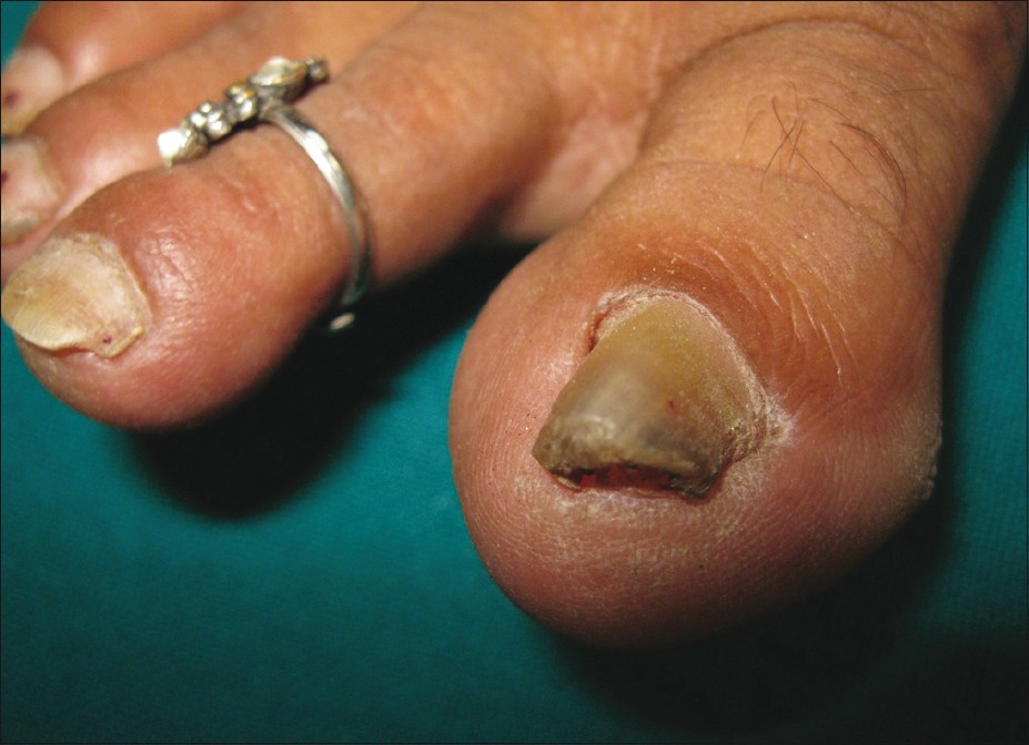
Figure 3: Pincer smash - Warts: It is the most common blast tumor, and mostly affects children and young adults. Periungual warts [Figure - 4] are usually due to HPV-1, 2 and 4. Development of periungual warts is favored past maceration and trauma, especially boom bitter. The natural grade of warts restricts ambitious approaches to selected cases. Partial or complete nail avulsion is indicated for exploring the extent of involvement of nail bed or matrix with HPV and as well to ensure complete eradication of diseased tissue. Medical treatments, usually topical, include keratolytic agents, virucidal agents and immunomodulators. All choices have been utilized successfully, only keratolytic agents are the best first-line approach. Surgical treatments include cryotherapy, surgical excision, electrosurgery, infrared coagulation, localized heating with a radiofrequency heat generator and light amplification by stimulated emission of radiation therapy, especially the Er: YAG laser. Recalcitrant periungual verrucae (24 lesions) in 17 patients were vaporized with the carbon dioxide laser in combination with partial or consummate boom avulsion. [24] A complete cure rate of 71% was observed in patients who had one or 2 treatments. The cure rate increased to 94% in patients who underwent one or 2 laser treatments in combination with other therapies. Postoperative pain was short lived and infection and significant onychodystrophy were uncommon.
-
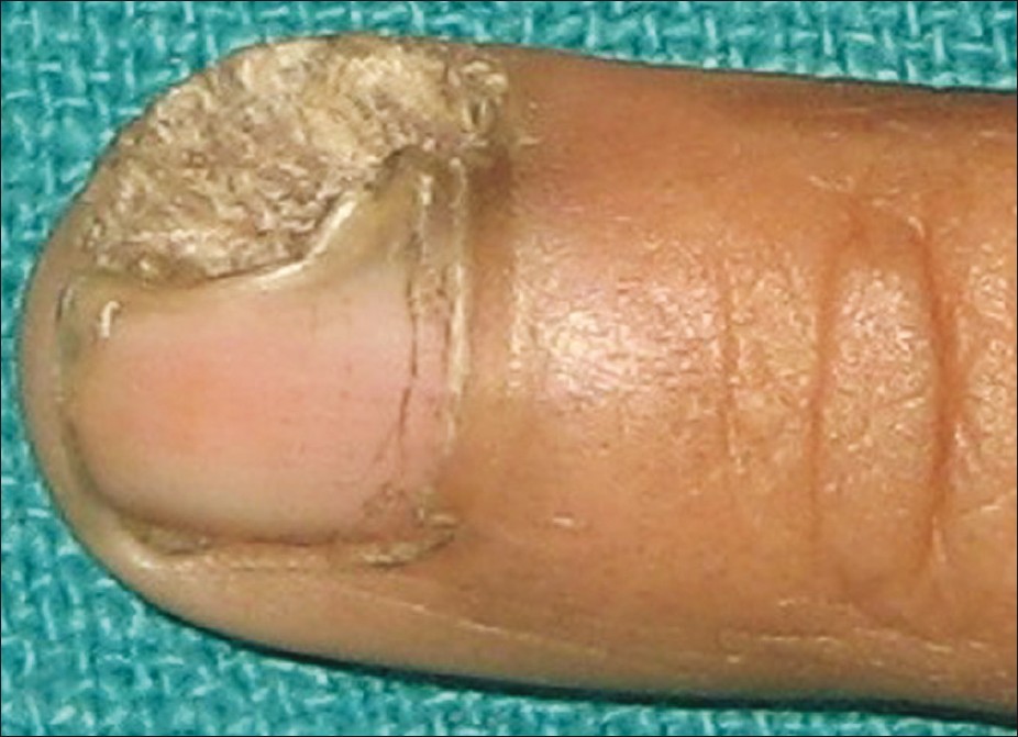
Effigy 4: Wart on the lateral nail fold encroaching on smash bed and matrix resulting in devastation of the boom plate. Partial nail avulsion is indicated to determine the extent of the wart - Tumors: Nail plate avulsion combined with nail bed excision forms the treatment for the following tumors:
- Onychomatricoma: Information technology is a rare nail matrix tumor with specific clinical and histologic features, including a macroscopic appearance of filiform digitations originating from the nail matrix that are inserted in the nail plate. [25] Onychomatricoma has a classical clinical appearance; however, information technology is difficult to place, equally it is not until surgery, when the typical filiform projections are more than visible that the diagnosis can exist made. [26]
- Glomus tumor [Effigy - 5]: Information technology is a painful subcutaneous nodule, commonly occurring in the subungual regions, and is accompanied past tenderness and temperature sensitivity. In the treatment of subungual glomus tumor, surgical excision is known to exist the only curative method. Information technology is challenging to minimize postoperative blast deformity and yet to ensure a depression incidence of tumor recurrence. [27] The transungual approach with smash avulsion and an incision selected according to the tumor location tin produce an splendid outcome with minimal postoperative complications. Dressing with a trimmed nail plate may also exist beneficial in managing the wound. [28]
- Miscellaneous: Melanoma and nonmelanoma cancers, pyogenic granuloma, fibrokeratoma and exostoses are the other tumors that may require a boom avulsion every bit a preliminary procedure. [3]
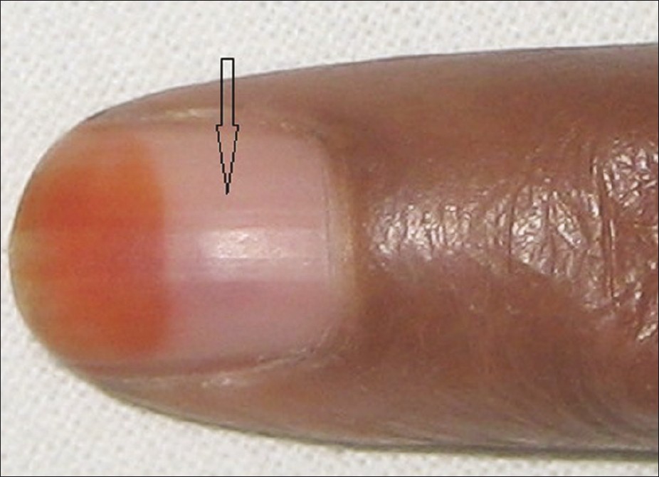
Figure v: Distal nail plate showing henna staining and intensely painful dusky erythematous lesion (arrow) under the nail plate, which was diagnosed every bit Glomus tumor
Contraindications
Relative contraindications to performing surgery in the nail unit are outlined as follows [3] :
- Peripheral vascular disease
- Collagen vascular disease
- Diabetes mellitus
- Disorders of hemostasis
- Astute infection or inflammation of the boom unit, including the surrounding paronychial tissues
Procedures of Nail Avulsion
Distal [iii],[29],[30],[31] and proximal avulsion are the two surgical approaches for undertaking nail avulsion. [29],[32] Chemical avulsion with urea paste is another valid nonsurgical technique that may be used in sure situations. A partial or complete boom avulsion can exist performed, depending on the location and extent of disease. Nail avulsion, however, is not a definitive cure in cases of blast dystrophy acquired by onychocryptosis, nail matrix illness [33] or all-encompassing nail bed pathology.
Anesthesia
Before avulsion, anesthesia of the digit is achieved through a digital cake performed with 1% lidocaine. [34]
There are 2 schools of idea on the use of epinephrine with lidocaine in the context of digital anesthesia. By convention, when administering an coldhearted for nail surgery, the utilize of epinephrine should be avoided, specially in patients with a history of all-encompassing vascular illness. Therefore, patients with a history of thrombotic or vasospastic illness and uncontrolled hypertension should not receive epinephrine. [3] Epinephrine has vasoconstricting properties, and it has been associated with necrosis and poor wound healing of tissues, while others believe that these complications appear to be more often than not theoretical and have rarely been noted to occur in practice. Proper injection technique and adequate choice of patients are recommended to minimize complications. A concentration of ane:two,00,000 is deemed as safe. Indeed, in a study, epinephrine-supplemented local anesthetics were used for the ear and nose surgery without whatsoever significant complications in more than than 10,000 surgical procedures. [35] Further, peel blood menses was studied at the fingerpads via laser Doppler flowmetry over the class of 24 h in a prospective, double-bullheaded, randomized, placebo-controlled written report with twenty vascularly healthy test persons. It was shown that adrenaline additive in local anesthesia decreased claret catamenia by less than 55% after a menstruum of 16 min. [36] In notwithstanding another study, in that location were 3110 consecutive cases of elective injection of depression-dose epinephrine (one:100,000 or less) in the hand and fingers and no instance of digital tissue loss was documented. [37] In fact, a total of fifty cases of digital gangrene have been documented in the literature, of which 21 were associated with the use of epinephrine; however, this was prior to 1950, when procaine was used as an anesthetic and the concentration of epinephrine used was high. The adrenaline digital infarction cases that created the dogma are invalid because they were likewise injected with either procaine or cocaine, which were both known to crusade digital infarction on their ain, and none of the 21 adrenaline infarction cases had an attempt at phentolamine rescue. [38] In fact, the addition of epinephrine reduces the need for the use of tourniquets and large volumes of anesthetic and provides a better and longer pain command during digital procedures. Exsanguinating tourniquet may be used to minimize bleeding; however, it should be released every xv min for a few minutes to preclude gangrene. [39]
Various methods have been suggested to reduce the pain while injecting a local anesthetic. Addition of sodium bicarbonate reduces the stinging sensation related to the acidic nature of adrenaline containing local coldhearted. Sedation with a combination of sedatives, analgesics and tranquilizers is helpful. This places the patient in a quiescent country and then that local anesthetic and nerve blocks may be comfortably administered. In add-on, repetitive, rapid pinching and shaking of the skin proximal to the site of injection during lignocaine infiltration works on the "gate control hypothesis." [40]
Methods
Nail avulsion surgery is oft accomplished using a nail elevator device. In addition, a mosquito hemostat or a dental spatula may also be used for the purpose. In distal nail avulsion [Effigy - half dozen]a-d, the instrument is introduced under the distal free border of the nail plate and so that the smash plate can exist separated from the underlying blast bed hyponychium. The nail plate is so separated from the underlying nail bed directed proximally towards the matrix, with pregnant resistance occurring until the matrix is reached. As the matrix is reached, the surgeon experiences a sudden decrease in resistance. Later, the elevator [Figure - 7] is reinserted with several longitudinal and side to side strokes to disassemble the nail plate from the nail bed totally. Thereafter, the elevator is inserted nether the PNF in the proximal nail groove betwixt the eponychium and the nail plate to release the attachment. This step should exist a gentle one so equally to avert inadvertent injury. [33]
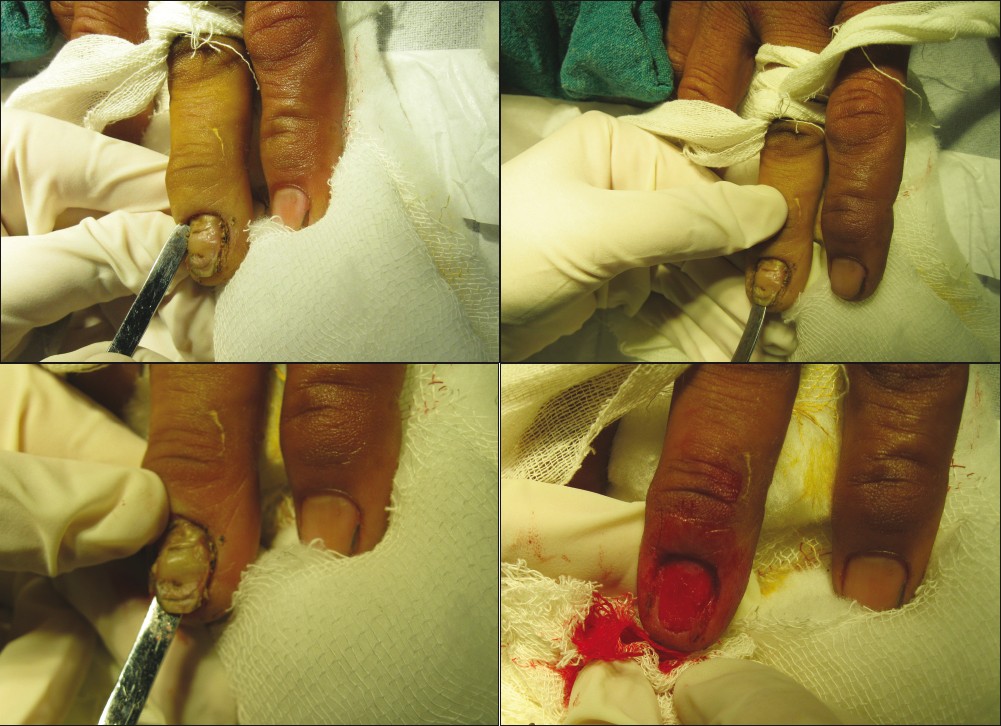 |
| Figure 6a: Distal smash avulsion in a case of chronic paronychia with proximal nail fold fibrosis and dystrophic nail plate. The plate is existence avulsed in addition to crescentic excision of the proximal nail fold Effigy 6b: Freeing of the lateral blast fold Figure 6c: Lifting of the smash plate from the nail bed with lateral sweeping movement Figure 6d: Vascular blast bed after separation of the smash plate |
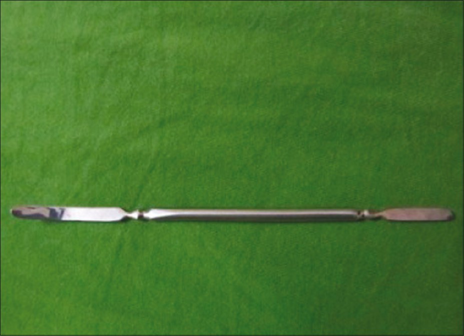 |
| Figure 7: Freer's elevator |
Proximal nail avulsion is preferred in the presence of distal nail dystrophy, in which it is often not possible to access the distal costless edge of the nail plate. This situation is often encountered in distal subungual onychomycosis. [29],[32],[41] The Freer elevator is inserted beneath the cuticle in the proximal groove to carve up the PNF from the blast plate. And then, it is reoriented so as to allow its concave surface to accommodate the curved surface of the ventral surface of the smash plate. [42] The instrument is advanced until it finally reaches the distal edge of the boom plate.
Secondary bacterial infections can be a cause of considerable morbidity in nail avulsion, especially in toe nails. Intraoperative antiseptic nail irrigation has, therefore, been recommended to reduce bacterial contagion. [43]
Alternatives to the aforementioned blast avulsion methods have been described. In many cases, partial nail plate avulsion is preferable compared with traditional full distal and proximal plate avulsions. The techniques described herein include partial distal, lateral, proximal and window techniques and two variations of the total plate avulsion termed the trap door and lateral nail plate ringlet avulsion. [44] Past using these methods, the surgeon is able to access the targeted blast unit while minimizing trauma to the next, uninvolved tissue.
Chemic boom avulsion
Forty percent urea ointment is often used in the treatment of onychomycosis, onychogryphosis, psoriasis and candidal and bacterial infections. [45],[46],[47] Urea ointment paste is formulated to include 40% urea, 5% white beeswax or paraffin, 20% anhydrous lanolin and 35% white petrolatum. [45],[46] Urea acts past dissolving the bond between the nail bed and the nail plate, and it too softens the smash plate. The paronychial area is protected with agglutinative tape earlier applying urea paste to prevent chemical irritation of the soft tissues. The ointment is liberally applied to the nail plate, and hypoallergenic tape is used to create a well around the treated thickened nail to hold the paste. [47] The patient is instructed to go on the nail occluded and to avoid wetting the treated expanse. After 1 week of apoplexy, the dystrophic nail is removed past using a nail elevator and a nail clipper. In onychomycosis, antifungals may be prescribed as an adjunct. Nonetheless, surgical nail avulsion followed by topical antifungal therapy cannot exist recommended for the treatment of onychomycosis every bit information technology is often associated with a high dropout rate and poor compliance. [12] Dystrophic nails may respond improve to chemical avulsion, and it is the platonic management for symptomatic dystrophic nails in patients with diabetic neuropathy, vascular disease or immunosuppression. [45] In improver, chemical avulsion may be used as a palliative, hurting-relieving therapy in onychogryphosis. Minimal to absent pain, a depression risk of infection, hemorrhage and bottom downtime are the advantages of chemic over surgical avulsion. [45] Requirement of prolonged application and irritation are some disadvantages of the procedure. Further, patients with gross thickening of the nail without significant blast dystrophy may reply poorly to chemical avulsion due to poor penetration. [45] Superficial abrasion of the nail plate may exist worthwhile to foster the penetration of urea. Contamination with water and poor occlusion may lead to treatment failure. Preparations other than 40% urea have besides been tried. A combination of 20% urea and 10% salicylic acid ointment under a 2-week occlusion has been finer used for minimally dystrophic nails. [30] Besides, a nail lacquer formulation containing 40% urea in a flick-forming solution has been devised. In a study comprising ten patients of onychomycosis, the urea nail lacquer was practical with a brush twice a day for 1 week by the patient and for a farther week in ii patients presenting with total dystrophic onychomycosis. This facilitated piece of cake removal of nail and was well tolerated. [48] In another report comprising of 13 patients of onychomycosis, a solution of 1% fluconazole and 20% urea in a mixture of ethanol and h2o applied once daily at bedtime showed a favorable response. [49] Phenolization is a well-conceived method of chemic matricectomy. In this procedure, 88% phenol is applied to the nail matrix while intendance is taken to avoid contamination with whatever ointment practical in the vicinity. It may be practical in iii cycles of 30 s each. Phenol has to be applied with the tourniquet on, and so as to ensure a bloodless field, as claret is known to inactivate phenol. Finally, phenol is neutralised with isopropyl booze and an appropriate dressing is done. [fifty] In a randomized study conducted on 148 ingrowing nails (grade 2-3) of 110 patients, 1-min phenol cauterization of the germinal matrix was institute to accept a better rubber profile than prolonged applications in the handling of ingrown nails. [51] Unpredictable tissue damage and prolonged healing time are the disadvantages of phenolization. Recently, the fractional avulsion of the affected edge and treatment of the germinal matrix for 1 min with 10% sodium hydroxide preceded by matrix curettage has been found to be an effective and safety treatment modality for ingrown toenails in people with diabetes. [52]
Sodium hydroxide is an alternative chemic agent that has been claimed to cause less tissue damage every bit compared to phenol. Matricectomy with 10% sodium hydroxide, either applied for 2 min or ane min combined with curettage, is as constructive in the treatment of ingrowing toenails with high success rates and minimal postoperative morbidity. [53] In a study comprising 46 patients, 154 ingrowing nail sides were treated with either sodium hydroxide or phenol matricectomy. Both sodium hydroxide and phenol were found to be effective giving loftier success rates, but sodium hydroxide caused less postoperative morbidity and provided faster recovery. [54]
Laser nail avulsion
The use of carbon dioxide laser has recently been well described. A 63-yr-old man was evaluated and treated with the carbon dioxide laser for a persistent pincer blast deformity. The patient tolerated the procedure well and had an acceptable surgical outcome. [21] In another study, 196 consecutive patients previously unsuccessfully treated past surgery underwent successful CO two light amplification by stimulated emission of radiation (v W, defocused ii mm beam in continuous mode) surgery for recurrent onychocryptosis. [55] Partial blast avulsion followed past matricectomy with pulse CO 2 laser in the treatment of ingrown toenails resulted in a loftier cure rate, short postoperative pain duration and low risk of infection. [56] Recalcitrant periungual verrucae (24 lesions) in 17 patients were successfully treated with CO 2 laser vaporization. [57] Vaporization of these warts, in combination with partial or complete nail avulsion, resulted in complete cures in 71% of the patients who underwent one or two treatments. Infection and significant onychodystrophy were uncommon. Pain was largely short lived. Thus, laser therapy in combination with nail avulsion improves the therapeutic outcome and reduces complications.
Postoperative care
Meticulous postoperative care is essential for a successful nail avulsion. A nonadherent, highly absorbant dressing is ideal. It may be kept in place with either an elastoplast or a newspaper record. The latter is less sticky and user-friendly to remove. The lateral grooves may exist studded with either a alkane gauze or an antibiotic tulle. [i],[3],[4] Dressing may be removed after 24 h afterward soaking in warm water or saline. It is an acceptable do to soak the operated area in warm water twice a day. In add-on, the povidone-iodine solution awarding may promote the healing procedure. The patient should be advised to continue the operated limb elevated so as to minimize the pain and swelling. Likewise, minimal activity with the involved limb, peculiarly if toenails are avulsed, should be carried out for at least 2 weeks.
Complications
Complications are seldom encountered in nail avulsion. These largely event from blast matrix impairment and present with postoperative smash deformity. Pain is the most common complexity following blast avulsion. Information technology is unremarkably of curt duration and responds well to analgesics. [58] Allergy to anesthetic, minor wound discharge, infection, hematoma, nail deformity, malalignment, blast impaction (distal embedding), local spicule growth and persistent hurting and swelling are the other adverse sequelae. Complications may be avoided past adequate preventive measures, such as judicious patient selection, aseptic technique and gentle handling of the nail matrix. [59]
Conclusion
Nail avulsion is a subject that has not been paid attention to by dermatologists. I needs to be well-versed with the anatomy of the nail while undertaking a nail avulsion to avoid matrix and nail fold injury. Total nail avulsion has been the conventional method to deal with various nail unit pathologies; even so, partial avulsion has gained popularity due to its simplicity and fewer postoperative complications. Ingrown toe nail, chronic onychomycosis and periungual warts go on to be the near common indications for blast avulsion. Conscientious patient selection and maintenance of asepsis during and later the procedure and gentle handling of matrix and nail folds are the key to superior outcomes of the procedure.
References
| 1. | Siegle RJ, Swanson NA. Nail surgery: A review. J Dermatol Surg Oncol 1982;8:659-66. [Google Scholar] |
| 2. | Haneke Due east. Surgical anatomy of the smash apparatus. Dermatol Clin 2006;24:291-6. [Google Scholar] |
| 3. | Scher RK. The nail. In: Roenigk RK, Roenigk HH, Ratz JL, editors. Dermatologic Surgery-Principles and Practice. 2 nd ed. New York: Marcel Dekker; 2006. p. 281-eight. [Google Scholar] |
| 4. | Clark RE, Madani S, Bettencourt MS. Nail surgery. Dermatol Clin 1998;16:145-64. [Google Scholar] |
| 5. | Zuber TJ. Ingrown toenail removal. Am Fam Physician 2002;65:2547-52. [Google Scholar] |
| six. | Rounding C, Bloomfield Due south. Surgical treatments for ingrowing toenails. Cochrane Database Syst Rev 2005;eighteen: CD001541. [Google Scholar] |
| vii. | Bos AM, van Tilburg MW, van Sorge AA, Klinkenbijl JH. Randomized clinical trial of surgical technique and local antibiotics for ingrowing toenail. Br J Surg 2007;94:292-6. [Google Scholar] |
| viii. | Gerritsma-Bleeker CL, Klaase JM, Geelkerken RH, Hermans J, van Det RJ. Partial matrix excision or segmental phenolization for ingrowing toenails. Arch Surg 2002;137:320-5. [Google Scholar] |
| 9. | Shaikh FM, Jafri 1000, Giri SK, Keane R. Efficacy of wedge resection with phenolization in the treatment of ingrowing toenails. J Am Podiatr Med Assoc 2008;98:118-22. [Google Scholar] |
| 10. | Rollman O. Handling of onychomycosis by partial blast avulsion and topical miconazole. Dermatologica 1982;165:54-61. [Google Scholar] |
| eleven. | Lai WY, Tang WY, Loo SK, Chan Y. Clinical characteristics and handling outcomes of patients undergoing nail avulsion surgery for dystrophic nails. Hong Kong Med J 2011;17:127-31. [Google Scholar] |
| 12. | Grover C, Bansal Due south, Nanda S, Reddy BS, Kumar 5. Combination of surgical avulsion and topical therapy for unmarried blast onychomycosis: A randomized controlled trial. Br J Dermatol 2007;157:364-eight. [Google Scholar] |
| xiii. | Flim-flam IM. Osteomyelitis of the distal phalanx following trauma to the smash: A instance report. J Amer Podiatric Assoc 1992;82:542-4. [Google Scholar] |
| 14. | Tucker DJ, Jules KT, Raymond F. Nailbed injuries with hallucal phalangeal fractures-evaluation and treatment. J Amer Podiatric Assoc 1996;86:170-3 [Google Scholar] |
| 15. | Banks As, Cain TD, Ruch JA. Physeal fractures of the distal phalanx of the hallux. J Am Podiatr Med Assoc 1988;78:310-3. [Google Scholar] |
| sixteen. | Hashizume H, Nishida G, Mizumoto D, Takagoshi H, Inoue H. Dorsally displaced epiphyseal fracture of the phalangeal base. J Hand Surg Br 1996;21:136-eight. [Google Scholar] |
| 17. | Grover C, Bansal S, Nanda S, Reddy BS, Kumar 5. En bloc excision of proximal nail fold for treatment of chronic paronychia. Dermatol Surg 2006;32:393-8. [Google Scholar] |
| xviii. | Bednar MS, Lane LB. Eponychial marsupialization and nail removal for surgical treatment of chronic paronychia. J Manus Surg Am 1991;16:314-7. [Google Scholar] |
| 19. | de Berker DA, Richert B, Duhard Due east, Piraccini BM, André J, Baran R. Retronychia: Proximal ingrowing of the smash plate. J Am Acad Dermatol 2008;58:978-83. [Google Scholar] |
| twenty. | el-Gammal Southward, Altmeyer P. Successful conservative therapy of pincer nail syndrome. Hautarzt 1993;44:535-7. [Google Scholar] |
| 21. | Lane JE, Peterson CM, Ratz JL. Avulsion and partial matricectomy with the carbon dioxide light amplification by stimulated emission of radiation for pincer nail deformity. Dermatol Surg. 2004;30:456-8. [Google Scholar] |
| 22. | Baran R, Haneke E, Richert B. Pincer nails: Definition and surgical treatment. Dermatol Surg 2001;27:261-6. [Google Scholar] |
| 23. | Ghaffarpour G, Tabaie SM, Ghaffarpour Thou. A new surgical technique for the correction of pincer-nail deformity: Combination of splint and nail bed cutting. Dermatol Surg 2010;36:2037-41. [Google Scholar] |
| 24. | Street ML, Roenigk RK. Recalcitrant periungual verrucae: The part of carbon dioxide laser vaporization. J Am Acad Dermatol 1990;23:115-20. [Google Scholar] |
| 25. | Goutos I, Furniss D, Smith GD. Onychomatricoma: An unusual case of ungual pathology: Instance report and review of the literature. J Plast Reconstr Aesthet Surg 2010;63:e54-seven. [Google Scholar] |
| 26. | Estrada-Chavez One thousand, Vega-Memije ME, Toussaint-Caire S, Rangel Fifty, Dominguez-Cherit J. Giant onychomatricoma: Report of ii cases with rare clinical presentation. Int J Dermatol 2007;46:634-6. [Google Scholar] |
| 27. | Song M, Ko HC, Kwon KS, Kim MB. Surgical treatment of subungual glomus tumor: A unique and simple method. Dermatol Surg 2009;35:786-91. [Google Scholar] |
| 28. | Moon SE, Won JH, Kwon OS, Kim JA. Subungual glomus tumor: Clinical manifestations and outcome of surgical treatment. J Dermatol 2004;31:993-7. [Google Scholar] |
| 29. | Siegle RJ, Swanson NA. Nail surgery: A review. J Dermatol Surg Oncol 1982;viii:659-66. [Google Scholar] |
| 30. | Zook EG, Baran R, Haneke Eastward, Dawber RPR. Nail surgery and traumatic abnormalities In: Baran R, Dawber RP, de Berker DA, Haneke E, Tosti A, editors. Diseases of the Nails and their Management iii rd ed. Uk: Blackwell Scientific discipline Ltd; 2001. p. 425-514. [Google Scholar] |
| 31. | Scher RK. Surgical avulsion of nail plates by a proximal to distal technique. J Dermatol Surg Oncol 1981;7:296-seven [Google Scholar] |
| 32. | Scher RK. Nail surgery. Clin Dermatol 1987;five:135-42. [Google Scholar] |
| 33. | Albom MJ. Avulsion of a nail plate. J Dermatol Surg Oncol 1977;3:34-5 [Google Scholar] |
| 34. | Denkler K. A comprehensive review of epinephrine in the finger: To practice or non to do. Plast Reconstr Surg 2001;108:114-24. [Google Scholar] |
| 35. | Häfner HM, Röcken One thousand, Breuninger H. Epinephrine-supplemented local anesthetics for ear and olfactory organ surgery: Clinical employ without complications in more than ten,000 surgical procedures. J Dtsch Dermatol Ges 2005;3:195-9. [Google Scholar] |
| 36. | Häfner HM, Schmid U, Moehrle 1000, Strölin A, Breuninger H. Changes in acral blood flux under local application of ropivacaine and lidocaine with and without an adrenaline additive: A double-blind, randomized, placebo-controlled study. Clin Hemorheol Microcirc 2008;38:279-88. [Google Scholar] |
| 37. | Lalonde D, Bong M, Benoit P, Sparkes G, Denkler G, Chang P. A multicenter prospective written report of 3,110 sequent cases of constituent epinephrine use in the fingers and hand: The Dalhousie Projection clinical phase. J Mitt Surg Am 2005;30:1061-7. [Google Scholar] |
| 38. | Thomson CJ, Lalonde DH, Denkler KA, Feicht AJ. A critical look at the testify for and confronting elective epinephrine use in the finger. Plast Reconstr Surg 2007;119:260-6. [Google Scholar] |
| 39. | Krunic AL, Wang LC, Soltani Thousand, Weitzul Southward, Taylor RS. Digital anesthesia with epinephrine: An old myth revisited. J Am Acad Dermatol 2004;51:755-9. [Google Scholar] |
| twoscore. | Mutalik S. How to make local anesthesia less painful? J Cut Aesth Surg 2008;1:37-8. [Google Scholar] |
| 41. | Jellinek NJ. Smash surgery: Practical tips and treatment options. Dermatol Ther 2007;20:68-74. [Google Scholar] |
| 42. | Clark RE, Madani Southward, Bettencourt MS. Nail surgery. Dermatol Clin 1998;16:145-64. [Google Scholar] |
| 43. | Becerro de Bengoa Vallejo R, Losa Iglesias ME, Cervera LA, Fernández DS, Prieto JP. Efficacy of intraoperative surgical irrigation with polihexanide and nitrofurazone in reducing bacterial load after nail removal surgery. J Am Acad Dermatol 2011;64:328-35. [Google Scholar] |
| 44. | Collins SC, Cordova K, Jellinek NJ. Alternatives to consummate nail plate avulsion. J Am Acad Dermatol 2008;59:619-26. [Google Scholar] |
| 45. | S DA, Farber EM. Urea ointment in the nonsurgical avulsion of smash dystrophies: A reappraisal. Cutis 1980;25:609-12. [Google Scholar] |
| 46. | White MI, Clayton YM. The treatment of fungus and yeast infections of nails by the method of "chemical removal'. Clin Exp Dermatol 1982;vii: 273-6. [Google Scholar] |
| 47. | Averill RW, Scher RK. Simplified smash taping with urea ointment for nonsurgical nail avulsion. Cutis 1986;38:231-3. [Google Scholar] |
| 48. | Baran R, Tosti A. Chemical avulsion with urea blast lacquer. J Dermatolog Treat 2002;13:161-iv. [Google Scholar] |
| 49. | Baran R, Coquard F. Combination of fluconazole and urea in a boom lacquer for treating onychomycosis. J Dermatolog Care for 2005;16:52-5. [Google Scholar] |
| fifty. | Bostanci Due south, Ekmekçi P, Gürgey Due east. Chemic matricectomy with phenol for the treatment of ingrowing toenail: A review of the literature and follow-up of 172 treated patients. Acta Derm Venereol 2001;81:181-iii. [Google Scholar] |
| 51. | Tatlican S, Yamangöktürk B, Eren C, Eskioðlu F, Adiyaman Due south. Comparison of phenol applications of different durations for the cauterization of the germinal matrix: An efficacy and safety written report. Acta Orthop Traumatol Turc 2009;43:298-302 [Google Scholar] |
| 52. | Tatlican South, Eren C, Yamangokturk B, Eskioglu F, Bostanci S. Chemic matricectomy with 10% sodium hydroxide for the treatment of ingrown toenails in people with diabetes. Dermatol Surg 2010;36:219-22. [Google Scholar] |
| 53. | Ozdemir E, Bostanci South, Ekmekci P, Gurgey Due east. Chemical matricectomy with 10% sodium hydroxide for the treatment of ingrowing toenails. Dermatol Surg 2004;30:26-31. [Google Scholar] |
| 54. | Bostanci S, Kocyigit P, Gurgey E. Comparing of phenol and sodium hydroxide chemical matricectomies for the treatment of ingrowing toenails. Dermatol Surg 2007;33:680-five. [Google Scholar] |
| 55. | Serour F. Recurrent ingrown large toenails are efficiently treated by CO2 laser. Dermatol Surg 2002;28:509-12 [Google Scholar] |
| 56. | Yang KC, Li YT. Treatment of recurrent ingrown keen toenail associated with granulation tissue by fractional nail avulsion followed by matricectomy with sharpulse carbon dioxide laser. Dermatol Surg 2002;28:419-21. [Google Scholar] |
| 57. | Street ML, Roenigk RK. Recalcitrant periungual verrucae: The role of carbon dioxide laser vaporization. J Am Acad Dermatol 1990;23:115-20 [Google Scholar] |
| 58. | Lai WY, Tang WY, Loo SK, Chan Y. Clinical characteristics and treatment outcomes of patients undergoing nail avulsion surgery for dystrophic nails. Hong Kong Med J 2011;17:127-31. [Google Scholar] |
| 59. | Moossavi M, Scher RK. Complications of nail surgery: A review of the literature. Dermatol Surg 2001;27:225-8. [Google Scholar] |
Source: https://ijdvl.com/nail-avulsion-indications-and-methods-surgical-nail-avulsion/
Posted by: donaldsonheiset.blogspot.com
0 Response to "What To Do For A Nail Avulsion"
Post a Comment